Rebuilding the Support Your Smile Needs
Bone Grafting
We understand how discouraging it can be to learn that jawbone loss might be standing in the way of the dental implants you need. Give our office a call to learn more about your options.
Your Strongest Smile Starts with a Solid Foundation
When a tooth is lost, the jawbone that once supported it naturally begins to shrink or recede. Over time, this can leave the area without enough healthy bone to securely anchor a dental implant. For many, this is where the journey to a restored smile seems to stop.
We believe that a successful, lasting restoration starts with an unshakeable foundation. Bone grafting isn’t just a procedure; it’s the essential first step we take to ensure your dental implants have the support they need to function beautifully for years to come. Our approach is built on precision, using state-of-the-art i-CAT 3D imaging to map out a treatment that is perfect for you.
Tailoring Your Treatment with Advanced Materials
You can think of it as our surgeons giving your body a helping hand. We carefully place an advanced, sterile grafting material in the precise spot where your jaw needs added strength and volume for an implant.
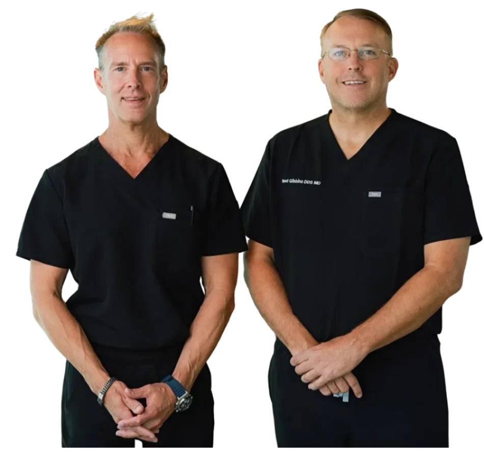
This is where the expertise of Dr. Draper and Dr. Gibbins comes in. They will recommend the precise material that gives you the surest path to a predictable and successful outcome. Your personalized plan will draw from several advanced options:
- Donor Materials (Allografts & Xenografts): These are highly processed, safe, and widely used materials that provide an excellent framework for your own bone to regenerate. They are a trusted and effective way to rebuild bone without needing a second surgical site.
- Synthetic Materials & Growth Factors: In some cases, a biocompatible synthetic material may be the best choice. We can also use Bone Morphogenetic Proteins (BMPs), which are powerful, naturally occurring proteins that act as a catalyst to accelerate your body’s own healing and bone formation.
Common Paths to a Stronger Jaw
Bone grafting is a versatile technique our surgeons use to solve several common issues. While each procedure is tailored to the patient, they all share the same goal: creating a healthy, stable base for your future smile.
Socket Preservation
The smartest time to address bone loss is before it even begins. Socket preservation is a proactive procedure we perform at the time of a tooth extraction. By immediately filling the empty socket with a bone graft, we prevent the surrounding bone and gum tissue from collapsing. This simple step preserves your jaw’s natural structure, making your future dental implant procedure much simpler and more successful.
Ridge Augmentation
If bone has already been lost, a ridge augmentation can rebuild it. During this procedure, we expertly place bone grafting material to restore the original height and width of your jawline. This not only creates the solid base needed for an implant but also helps restore the natural contours of your gums and smile.
Sinus Lift
The maxillary sinuses are natural air spaces located just above your upper back teeth. When these teeth are lost, only a thin wall of bone may separate the sinus from your mouth, leaving insufficient room for an implant. In a sinus lift, we gently elevate the sinus membrane and place a bone graft beneath it. This common and highly successful procedure creates the strong, stable foundation required for upper dental implants.
Start Your Journey with a Clear Plan
Feeling uncertain about your options is normal, but you don’t have to navigate this alone. The first step is a personal consultation with our surgical team. We’ll sit down with you, listen to your goals, and use advanced 3D imaging to get a complete picture of your oral health.
Our commitment is to give you a clear, honest assessment and a treatment plan you can feel confident about.
Ready to find out if bone grafting can put dental implants back on the table for you? Give our Forney or Fate office a call to schedule your consultation.